Dechra

Home / Dogs / Pruritus / Partial or No Response / 4 Pillars of Atopy Treatment

4 Pillars of Atopy Treatment

Topical Therapy and Skin Anatomy

A Brief Review of Skin Anatomy

Familiarity with basic dermal anatomy aids in understanding how to best treat conditions involving this important organ. The skin is divided into 3 distinct layers: the hypodermis (subcutis), the dermis and the epidermis.
The hypodermis is the insulation layer and is composed mostly of fat, vasculature and glands. The dermis comprises hair follicles and sebaceous glands. lt is an integral part of the connective tissue system and provides tensile strength and elasticity for the skin. Cell growth, proliferation, adhesion, migration and wound healing all begin in the dermis.
The epidermis is the outermost layer. lt is composed of multiple layers of cells that are defined by their position and morphology. In this layer, 85% of the cells are keratinocytes (corneocytes). The epidermis is in contact with the environment and consists of 4 distinct layers: the stratum basale (innermost), stratum spinosum, stratum granulosum and stratum corneum (outermost).21
The stratum corneum is the most important layer for barrier function. Structurally, it is a layer of cornified keratinocytes (corneocytes) bound together by an extracellular lipid matrix composed of free fatty acids, cholesterol and ceramides. The keratinocytes and extracellular matrix form a “brick-and-mortar” system which maintains the skin barrier, preventing invasion of allergens and infectious agents.18,21 Ceramides in particular play a crucial role in this barrier, representing 50% of the lipids in the stratum corneum. They are composed of a fatty acid and sphingoid base. One type of sphingoid base is phytosphingosine.9 When the stratum corneum is compromised, skin inflammation and irritation occur as allergens, bacteria and fungi penetrate the broken barrier.
Two electron microscopy studies report similar findings of defective epidermal lipid barrier in atopic dogs.8,16 ln atopic dogs, the deposition of lipid is more heterogeneous in the stratum corneum compared to healthy controls. Many areas in the intercorneocyte spaces lack lipids or often exhibit an abnormal lipid structure. ln the healthy canine stratum corneum, lipids are well arranged in compact sheets and fill in the space between cell layers.
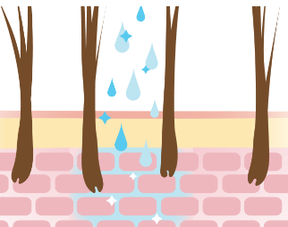
Figure. The normal epidermal barrier of a dog. The diagram shows the corneocytes and lipids working together to form a “brick-and-mortar” barrier.

Advantages of Topical Therapy19

These protocols have been suggested based on provided literature and are not intended to be “one size fits all.” They should be adjusted according to the individual needs of each patient.